Scanning for your Future
An MRI is painless, non-invasive and free of radiation. Because detailed images can be acquired from any angle, MRI is excellent in detecting most malignancies and neurological diseases of the brain, spine and pelvis. It’s also widely used with sports-related injuries, especially those affecting the knee, shoulder, hip, elbow and wrist. Exams of the organs of the chest and abdomen, can identify tumors and functional disorders.
Common MRI Studies
- Brain
- Spine
- Liver
- Male and female reproductive organs
- Pancreas
- Other soft tissues
Your MRI Exam
Because an MRI uses a strong magnetic field, advise us if you have a pacemaker, artificial heart valve, aneurysm clips, cochlear implants, a neurostimulator, metal pins/ plates/implants, foreign metal objects in your eye, an implanted drug infusion device, or if you are pregnant.
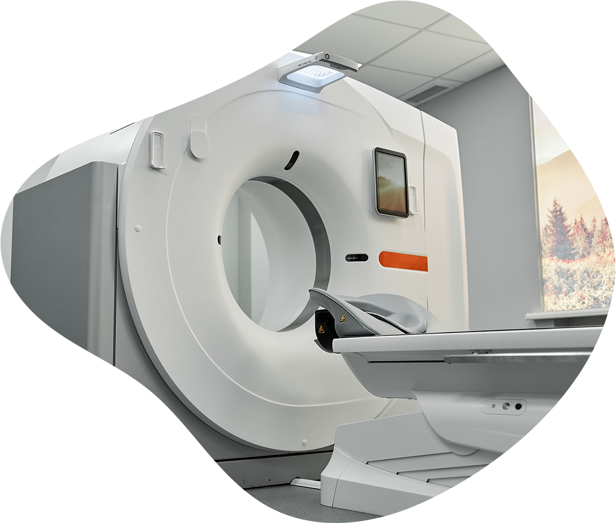
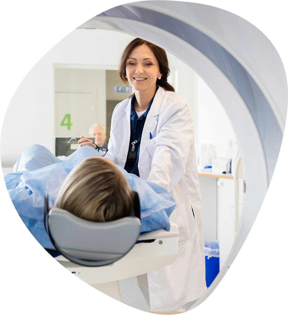
An MRI requires no special preparation. During the exam, you will lie on a comfortable table that will slide on a track into the scanner. A special coil may be used which will be placed around the area of your body to be scanned and will help isolate the imaging procedure.
If Magnetic Resonance Angiography (MRA) has been ordered by your physician, a contrast material may be administered through an IV in your arm. MRA is used to highlight certain blood vessels in the body to identify abnormalities such as aneurysms.
MR Arthrography is the study of a joint, such as the shoulder, knee, or wrist in which contrast material is placed into the joint. Once the contrast material is in place, images of the joint are obtained. The images are then evaluated by a radiologist for cartilage, ligament, and tendon problems in and around the joint. MR arthrography is more accurate and provides more information than a conventional MRI scan in many instances involving joints.


What to Expect for an MRI
Once an MRI exam begins, a technologist will be able to talk to you through a microphone on the outside of the exam room. While the MRI scanner is running, you will hear several loud knocking sounds. We will provide you with earphones for listening to music during the exam to make you more comfortable. Most MRI scans take between 30 – 45 minutes.
Next Steps:
We’ll report the results
A board certified radiologist will provide your physician with an interpretation of the MRI results.
Your Care Team will report to you
Your physician can then combine these results with your history and explain the findings to you.
We’re Here for You
It’s our specialty to take care of you and your health in times where imaging can be scary. We want everyone to feel welcome and comfortable in every process we provide. Reach out today to get started with our team!

